


Raman Microscopy for the Amateur Mineralogist.
| This page and the linked documents describe the construction, adjustment and use of a Raman microscope for the determination of minerals in rock sections. The availability of high performance laser cutoff filters and CCD detectors allows the development of such instruments with relatively few components at an affordable cost.
|
 |
Technical description of the Raman Microscope with first version of spectrograph.Page 1: Optical arrangement schematic. Page 2: Details of the design. Page 3: Beam splitter and filter. Page 4: First results and calibration. Page 5: References
|
||||||||||||||||||||||||||||||||||||||||||||
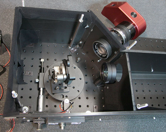 |
Raman Microscope version 2 with home build spectrograph.Improved performances of the spectrograph: wider spectral range, less field curvature... Page 6: design of the spectrograph. Page 7: Images and part lists. Page 8: tests, neon spectra. Page 9: tests, plastic spectra. Page 10: tests, the OH band.
|
||||||||||||||||||||||||||||||||||||||||||||
|
|
Raman microscope version 3 with double lasers: 532 nm and 632.8 nm.Page 11: first design with a small laser pointer. Page 12: addition of a 532nm laser design. Page 13: Examples of Raman spectra with 532nm laser, comparison with He-Ne laser
|
||||||||||||||||||||||||||||||||||||||||||||
|
|
Raman Microscope version 4 with triple lasers: addition of a 780 nm laser.Page 14: laser diode tests. Page 15: construction of 780 nm laser head Page 16: Raman spectra examples at 780nm
|
||||||||||||||||||||||||||||||||||||||||||||
|
|
|||||||||||||||||||||||||||||||||||||||||||||
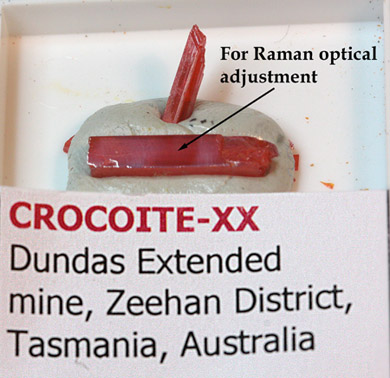
Raman spectra of mineral single crystals.The following pages show Raman spectra recorded with the system described above (old and new spectrograph) on single crystals. The spectra are compared to data from databases published on the web. Spectra recorded with the new spectrometer are marked with an asterisk. |
|||||||||||||||||||||||||||||||||||||||||||||
|
|||||||||||||||||||||||||||||||||||||||||||||