The Becke line and dark field methods
 |
 |
|
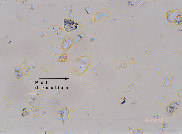
Figure 1.Becke line of quartz, liquid n=1.544. |
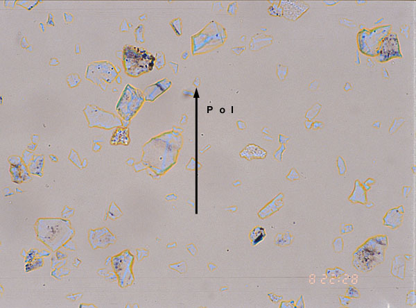
Figure 2. Idem with rotation of 90° of the polariser |
 |
 |
|
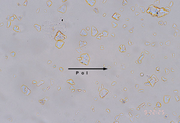
Figure 3. Becke line of quartz in liquid of n=1.553. |
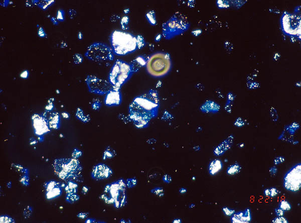
Figure 4. Dark field image of quartz in liquid of n=1.544. |
The Becke line method is usually utilized to determine if the crystal has a higher or lower index of refraction than the surrounding medium. The liquid used in figure 1 has the same index of refraction than the solid for a wavelength in the yellow part of the visible spectrum. The crystals appear thus with an orange rim (see theoretical explanation). The polariser of the central crystal is parallel to the small index of the crystal (ordinary index). In the top right image (figure 2), the polariser has been rotated by an angle of 90°and is now parallel to the high index of the crystal. The crossing point between the dispersion curves of liquid and solid has moved to the green region of the spectrum. The edges of the central crystal are slightly brighter as can be seen on the enlarged pictures (click on images to enlarge). If the index of refraction is increased to n= 1.553, the edges of the quartz particles become darker orange as the crossing point move to the red (see figure 3). The differences between the hue of crystal edges are less prominent than the ones observed with the phase contrast technique although the contrast of the images presented above has been slightly magnified by software to allow the reader to see differences between pictures.
The dark field technique is also illustrated. With the dark field attatchement of my Olympus microscope, only the blue edge of the crystal can be seen (figure 4). Some faint orange colors also appeared in some particles but too pale to be properly recorded on the photograph. Although this technique is well described in the literature, I have not pursued in that direction due to the difficulty to see the color effects with small variations of indices of refraction.
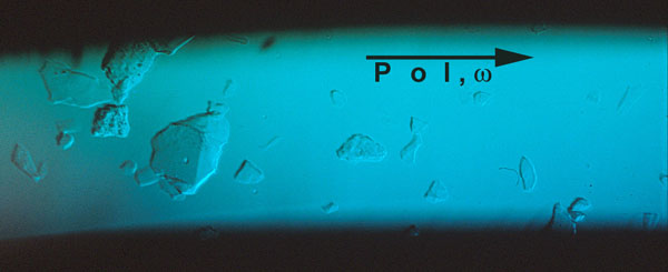
Fig.5 |
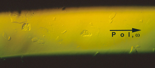
Fig.6 |
In the two figures above recorded with bright field illumination, the exit slit of the monochromator is in place so that only a small spectral range illuminates the stage of the microscope. In figure 5, the liquid has a higher index than the two crystals at the left which are thus visible. These two crystals disappear on the right picture because in the yellow light the small index of quartz is precisely equal to the liquid index. The technique can be used for the measurement of the index of refraction of the particles (determination of dispersion curves crossing points) but the tuning is less sharp than with phase contrast.