Conoscopy of Biaxial Minerals (2).
Figures normal to the acute bisectrix.
This page collects images of biaxial conoscopy as a function of the thickness of the crystal plate.
Video of the rotation of a muscovite conoscopic image perpendicular to the acute bisectrix.
Same muscovite but double thickness. Higher order isochromes are visible.
A single sheet of muscovite with a Benford plate. As usual, the isogyres are canceled and the rotation of the poles of the optic axes are visible.
Images below were obtained with muscovite crystals. The thickness of the plates increases from the top of the page to the bottom.
|
Figure 1a. Very thin plate with optic plane horizontal. Only the isogyres can be seen. No isochrome can be seen in the field of view due to the low birefringence. |
Figure 1 b. Rotation of the microscope stage at 45°. The isogyres are now hyperboles. |
Figure 1c. Same orientation as figure 1a but with a Benford plate inserted. Isogyres are removed, only the poles of the optic axes are visibles. |
|
Figure 2a. Thicker plate than in figure 1. Optic plane horizontal. Thinner isogyres close to the optic axes. The first isochromes visible are part of a lemniscate. |
Figure 2b. Hyperbolic isogyres at 45° orientation. The isochromes simply rotate as the crystal. |
Figure 2c. Same as 2a with Benford plate. Optic axes clearly visible. |


|
Figure 3a. O° orientation. Increased thickness. The vertical part of the isogyres becomes wider. First order isochrome is well developed, second order appears at the top and bottom of the image. |
Figure 3b. Orientation 45° with Benford plate inserted. The first order lemniscate is nearly complete. |


|
Figure 3c. Monochromatic light (467 nm). 45° with Benford plate. This figure and the following one show the influence of the wavelength of the light on the shape of the lemniscate moving around the poles of the optic axes. The second order extinction becomes visible at this wavelength near the edges of the figure. |
Figure 3d. Monochromatic light (628 nm). 45° with Benford plate. At this wavelength, lemniscate is wider. Second order is not visible in the field. |


|
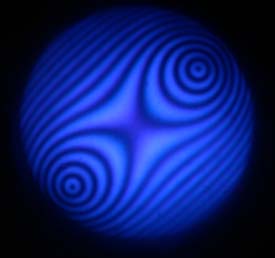
Figure 4a. O° orientation. Increased thickness. Monochromatic light 467 nm. Increased number of extinction curves. Closed curves around the optic axes appear.
|
Figure 4b. Orientation at 45°.
|
Figure 4c . O° orientation with Benford plate. If the stage is rotated with the Benford plate installed, the figure simply rotates with no changes. |
|
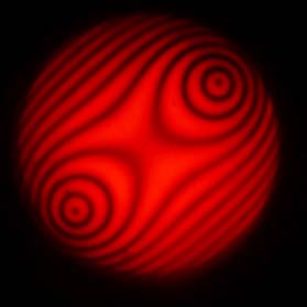
Figure 4d. Same image as figure 4a with wavelength equal to 628 nm. |
Figure 4e.Same image as figure 4b with wavelength equal to 628 nm. |
Figure 4f. Same image as figure 4c with wavelength equal to 628 nm. Only one closed curve is present at this wavelength around optic axes poles. |


|
Figure 5a. The highest thickness of the page. Vertical part of the isogyres is very wide. High birefringence colors present. |
Figure 5b. 45° orientation with Benford plate. The shape of lemniscates is clearly demonstrated. |
Figure 5c. 0° orientation. Wavelength = 467 nm. |
|
Figure 5d. 45° orientation. Good view of the hyperbolic isogyres. |
Figure 5e. 45° orientation with Benford plate. Very sharp poles of the optic axes. Lemniscates of high orders are visibles. |
Figure 5f. 45° orientation with Benford plate. Fewer isochromes than the blue image. |


Two superposed sheets of muscovite rotated by 90° from each other. This gives this amazing conoscopic figure from two indices ellipsoid in quadrature.
Next page [Conoscopy main page] [Home]